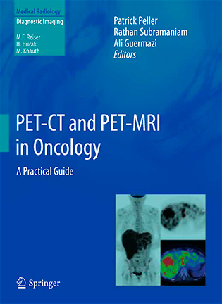
PET-CT and PET-MRI in Oncology - Patrick Peller
 Год выпуска: 2012
Автор: Patrick Peller, Rathan Subramaniam, Ali Guermazi
Жанр: Книги на английском
Формат: PDF
Качество: OCR
Описание: В этой книге рассматриваются различные аспекты ПЭТ / КТ и ПЭТ / МРТ в онкологии. Когда мы начали планировать книгу, мы стремились дать читателю четкое понимание физики, лежащей в основе этих очень сложных технологий, прежде чем описывать, как сканеры применяются в клинической онкологии. Мы начнем с главы, подробно объясняющей, как работают ПЭТ / КТ и ПЭТ / МРТ-сканеры и как максимизировать их присущие качества для достижения оптимальных результатов. Следующая глава дает довольно подробный обзор ПЭТ радиохимии и радиотрексеров. В третьей главе шаг за шагом вводятся основы интерпретации результатов ПЭТ / КТ и ПЭТ / МРТ. Мы считаем, что эти три вводные главы предоставят читателям прочную основу для понимания технологии, лежащей в основе этих методов визуализации. Это понимание абсолютно необходимо для интерпретации изображений, которые действительно могут быть очень сложными, и оно обеспечивает основу для центра книги, 14 глав, посвященных ПЭТ / Компьютерная Томография и ПЭТ / Магнитно Резонансная Томография в Киеве, применительно к наиболее часто встречающимся видам рака, включая проблемы с ВИЧ. Значительная роль ПЭТ / КТ в планировании лучевой терапии также включена. Эти главы щедро иллюстрированы. В последней главе теоретическое понимание первых трех глав и практические аспекты различных онкологических ситуаций объединены с подробной и тщательной главой об общих ошибках, связанных с использованием ПЭТ / КТ и ПЭТ / МРТ. Это мощные, но очень сложные технологии, и путь к диагностике усеян подводными камнями и артефактами, которые могут легко привести читателя к неправильному диагнозу. Знание этих потенциальных проблем сэкономит время и деньги, а также улучшит уход за больным.
Все авторы, которые участвовали в этой работе, являются известными экспертами в своих областях, из Северной Америки, Европы, Азии и Австралии. Мы в долгу перед ними за их работу и преданность делу и надеемся, что книга оправдает их лучшие ожидания.
Мы хотели бы посвятить эту книгу нашим женам Марибет, Сакиле и Ноуре и детям Синтии, Катрине, Джону, Мире, Анжане, Сантии, Дорре, Элиасу и Манелю. Без их терпения и любви эта книга, безусловно, не будет опубликована сегодня.
This book deals with various aspects of PET/CT and PET/MRI in oncology. When we began to plan the book, we aimed to give the reader a clear understanding of the physics that underlie these very complex technologies, before describing how the scanners are applied in clinical oncology. We begin with a chapter explaining in some detail how PET/CT and PET/MRI scanners work and how to maximize their inherent qualities for optimum results. The next chapter gives a fairly detailed overview of PET radiochemistry and radiotracers. The third chapter introduces, step by step, the basics of interpreting PET/CT and PET/MRI results. We think these three introductory chapters will provide readers with a strong basis for understanding the technology behind these imaging modalities. This understanding is absolutely necessary to interpret images that can be very complicated indeed, and it provides a foundation for the center of the book, 14 chapters on PET/CT and PET/MRI as applied to the most commonly encountered cancers including HIV issues. The significant role of PET/CT in planning radiotherapy is also included. These chapters are generously illustrated. In the final chapter, the theoretical understanding of the first three chapters and the practical aspects of various oncological situations are brought together with a detailed and thorough chapter on common errors associated with the use of PET/CT and PET/MRI. These are powerful but very complex technologies, and the path to diagnosis is strewn with pitfalls and artifacts that can easily lead the reader to the wrong diagnosis. Foreknowledge of these potential problems will save time and money, and will improve patient care.
All of the authors who participated in this work are renowned experts in their fields, from North America, Europe, Asia and Australia. We are indebted to them for their work and dedication and we hope the book will meet their best expectations.
We would like to dedicate this book to our wives Maribeth, Sakila and Noura and children Cynthia, Katrina, John, Meera, Anjana, Santhiya, Dorra, Elias and Manel. Without their patience and love, this book will certainly will not be public today.
Contents
«PET-CT and PET-MRI in Oncology: A Practical Guide»
Basics
- PET Physics and Instrumentation - Brad Kemp
- PET Radiochemistry and Radiopharmacy - Mark S. Jacobson, Raymond A. Steichen, and Patrick J. Peller
- PET/CT Interpretation - Patrick J. Peller
Oncologic Applications
- Central Nervous System - Jeffrey A. Miller and Terence Z. Wong
- Head and Neck - R. M. Subramaniam, J. M. Davison, U. Parikh, and M. Abou-Zied
- Chest - R. M. Subramaniam, J. M. Davison, D. S. Surasi, G. Russo, and P. J. Peller
- Breast - Gustavo A. Mercier, Felix-Nicolas Roy, and Francois Benard
- Gastrointestinal - Roland Hustinx
- Genitourinary - Jacqueline Brunetti and Patrick J. Peller
- Gynecologic - Patrick J. Peller
- Musculoskeletal - Jeffrey J. Peterson
- Hematology - R. M. Subramaniam, Leonne Prompers, A. Agarwal, Ali Guermazi, and Felix M. Mottaghy
- Dermatological - David Brandon and Bruce Barron
- Pediatric - Hossein Jadvar, Frederic H. Fahey, and Barry L. Shulkin
- Assessment of Response to Therapy - Ali Gholamrezanezhad, Alin Chirindel, and Rathan Subramaniam
- Metastatic Disease - Patrick J. Peller
- Radiotherapy Planning - Minh Tam Truong and Rathan M. Subramaniam
- Patients with HIV - R. M. Subramaniam, J. M. Davison, D. S. Surasi, T. Jackson, and T. Cooley
- Pitfalls and Artifacts - Geoffrey Bates Johnson and Christopher Harker Hunt
купить книгу: «PET-CT and PET-MRI in Oncology»
